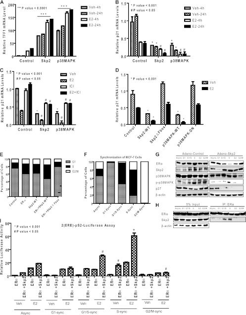
Fig 6.
Skp2 regulates ERα target gene expression, and the two proteins cooperate in driving S-phase entry of MCF-7 cells. (A to C) Real-time quantitative PCR analysis of TFF1 mRNA (A) or p21 mRNA (B) in MCF-7 cells infected with the control, Skp2, or p38MAPK adenovirus for 24 h followed by control, vehicle, or estradiol (E2) (10 nM) treatment for 4 h or 24 h and reversal of the E2 effect by the antiestrogen (1 μM) ICI 182,780 (C). (D) Real-time RT-PCR analysis of p21 mRNA in MCF-7 cells infected with control, Skp2-WT, Skp2ΔFbox, p38MAPK-WT, or p38MAPK-DN adenovirus for 24 h, followed by vehicle or ligand treatment. (E) FACS analysis. MCF-7 cells were infected with the control, ERα alone, Skp2 alone, or ERα and Skp2 adenovirus followed by 48 h of E2 (10 nM) treatment and propidium iodide staining to monitor the DNA content. (F) FACS analysis. MCF-7 cells were synchronized at various stages in the cell cycle, as shown in Fig. S5H in the supplemental material, and the percentages of cells in the different cell cycle stages in asynchronous (Async) and synchronized (sync) populations are shown. (G) Western analysis of the indicated proteins in MCF-7 cells synchronized at various stages of the cell cycle and infected with the control or Skp2 adenovirus. (H) Skp2 interacts with ERα preferentially in the G1/S and S phases of the cell cycle, as observed for synchronized MCF-7 cell populations. (I) Luciferase assay. Synchronized MCF-7 cells were transfected with the (ERE)2-pS2-Luciferase expression plasmid along with ERα and β-galactosidase, treated for 24 h with either the vehicle or E2 (10 nM), and monitored for luciferase activity. Data are from 3 experiments and are represented as means ± standard deviations (SD).

