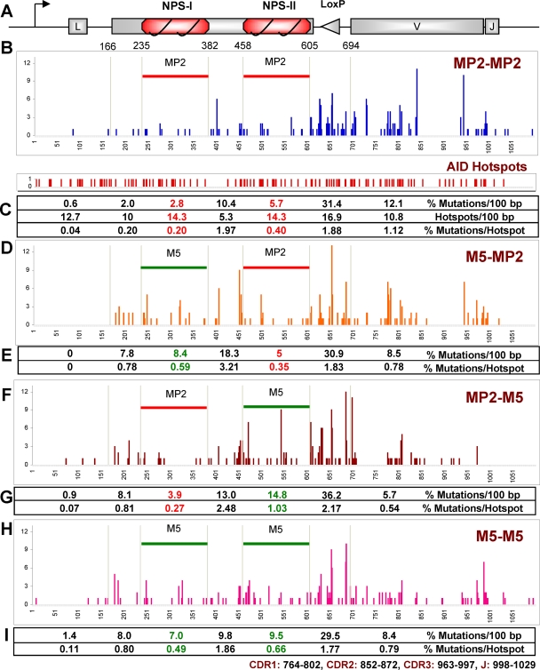
Fig 6.
Ig light chain sequence analysis of the nucleosome positioning sequence knock-in clones. (A) Map of lg gene with 2 MP2s (not to scale); the triangle represents the two recombined loxP sites. (B, D, F, and H) Mutations in 1.1 kb from the start of transcription (= 1); the numbers on the y axis represent point mutations at the indicated positions in MP2-MP2 (B), M5-MP2 (D), MP2-M5 (F), and M5-M5 (H). 1 to 165, IgL gene containing the leader region; 235 to 382, first NPS (MP2/M5); 383 to 457, spacer between two NPSs; 458 to 605, second NPS; 606 to 695, loxP site generated from the Bsr marker excision; 695 to 1100, IgL gene containing V and J regions. (C, E, G, and I) Summary of mutations. CDR, complementarity determining region.

