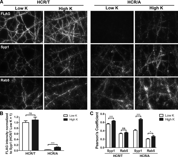
Fig 1.
HCR/T and HCR/A entry into cortical neurons. FLAG-HCR/T or FLAG-HCR/A (40 nM) was incubated with rat cortical neurons in low K buffer or high K buffer for 5 min at 37°C. (A) IF was performed to detect HCRs, Rab5, and synaptophysin 1 (Syp1). (B) Six random fields were selected for IF intensity analysis. The IF ratio between anti-FLAG and antisynaptophysin was used as a marker for HCR/T and HCR/A internalization. (C) HCR colocalization with Syp1 or Rab5 was analyzed as described in Materials and Methods. Student's t test was performed. ns, not significant (P > 0.05); *, P < 0.05; ***, P < 0.001. Error bars indicate standard deviations.

