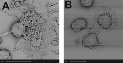
Fig 5.
Scanning electron photomicrographs of METs formed by bovine macrophages in response to M. haemolytica cells. Bovine macrophages (2.5 × 10⁵) were incubated with 5 × 10⁷ M. haemolytica cells or RPMI 1640 (negative control) for 5 min at 37°C. Cells were washed, fixed, and processed for SEM as described in Materials and Methods. (A) A matrix of extracellular DNA strands released in response to M. haemolytica cells (arrows indicate trapped bacterial cells); (B) control bovine macrophages incubated with RPMI that do not exhibit extracellular DNA fibrils. Photomicrographs are of representative cells from 3 independent experiments.

