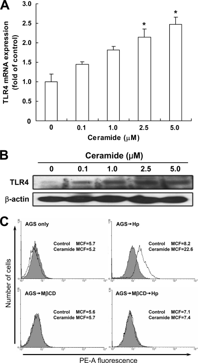
Fig 6.
H. pylori activates ceramide release in lipid rafts. AGS cells were treated with various concentrations of ceramide for 24 h. Total RNA and cell lysates were prepared to measure TLR4 mRNA and protein expression levels by quantitative real-time PCR (A) and Western blot analysis (B), respectively. The mRNA and protein expression levels were normalized to GAPDH and β-actin, respectively. (C) AGS cells were pretreated with medium alone or 5 mM MβCD for 30 min and then incubated with H. pylori at an MOI of 100 for 6 h. Cells were then stained with anti-ceramide (white histograms) or isotype control IgM (gray histograms), followed by Alexa Fluor 568-conjugated goat anti-mouse IgM, and the fluorescence intensity was assessed by flow cytometry. Results are expressed as means ± standard deviations. *, P < 0.05 compared with untreated control. MCF, mean channel fluorescence; MβCD, methyl-β-cyclodextrin.

