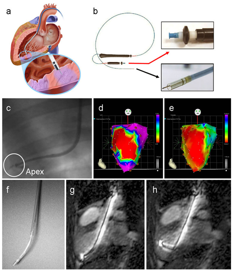
Fig. 1.
Imaging navigation of transendocardial delivery. a Schematic displays an injection catheter passed across the aortic valve into the left ventricular cavity, with its needle tip extruded into the endomyocardium of the inferoposterior wall (inset). Purple areas of myocardium highlight two injection sites (image kindly provided by Biologics Delivery Systems Group, Cordis Corporation). b MyoStar™ injection catheter. The black arrow points to an inset of the catheter tip with extended 27 gauge needle and the red arrow to the adjustable injector thumb knob and Luer-lock fitting for connection to the injection syringe. c Conventional X-ray fluoroscopic guidance of catheter positioning near the left ventricular apex (adapted with permission from [98]). d, e Examples of electromechanical mapping images in a patient with anterior myocardial infarction, with linear local shortening (d) and unipolar voltage (e) maps shown in the anteroposterior projection. The red areas correspond to the infarct territory, with coupling of the reduced electrical signal and mechanical function over the anterior left ventricular wall. In this case, intramyocardial injections of cell therapy were administered in a peri-infarct distribution, as indicated by the brown circular markers (reproduced with permission from [33]). f–h MR guidance of catheter-based injection. The flexibility of the custom MR-compatible injection catheter (f) enables its manipulation to all regions of the endocardial surface. (g, h) Active catheters generate a high signal intensity for easy visualisation inside the ventricular cavity using real-time MR steady-state free precession imaging (reproduced with permission from [162])

