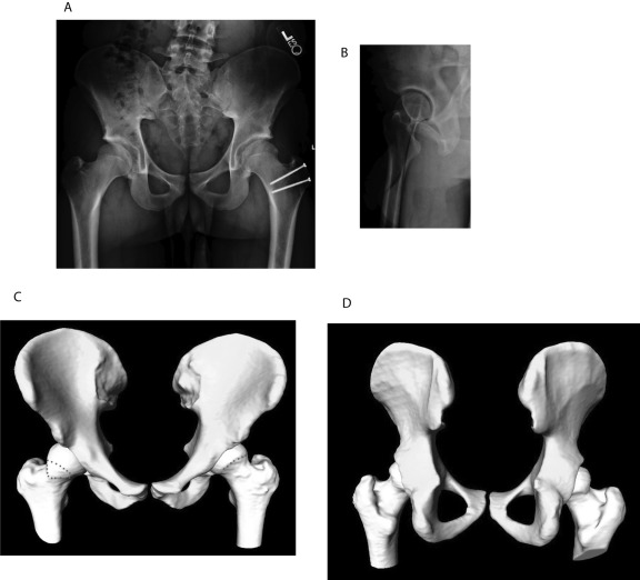
Fig. 14.
Case 6. A: Anteroposterior pelvic radiograph illustrating acetabular retroversion and a positive posterior wall sign in the right hip. B: False-profile lateral radiograph of the right hip, illustrating anterior acetabular overcoverage. C: Three-dimensional computational model confirming acetabular retroversion of the right hip with a reduced femoral head-neck offset. The red dotted lines on the right hip show the area that has reduced head-neck offset. The red dotted line on the left hip shows the new head-neck contour. Postsurgical correction of femoral head-neck offset is confirmed in the left hip. D: Three-dimensional computational model demonstrating the posterior wall deficiency of the left hip.

