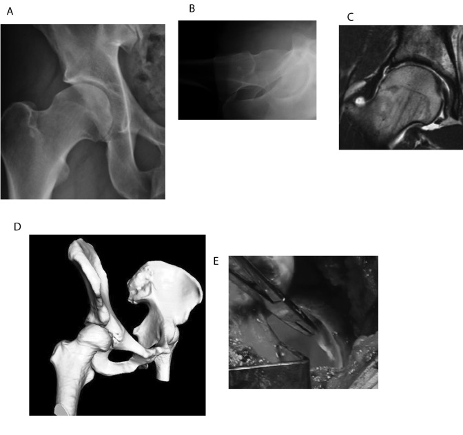
Fig. 17.
Case 9. A: Anteroposterior radiograph of the right hip, demonstrating acetabular retroversion and reduced femoral head-neck offset. B: Cross-table lateral radiograph demonstrating reduced femoral head-neck offset. C: T1-weighted magnetic resonance arthrogram demonstrating hyaline cartilage delamination. D: Three-dimensional computational model confirming acetabular retroversion and reduced femoral head-neck offset. E: Intraoperative photograph illustrating acetabular hyaline cartilage delamination from the one o'clock position to the three o'clock position.

