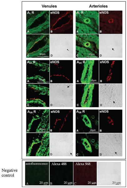
Figure 5.
Immunofluorescent assay demonstrates the presence of ADO A1, A2A, and A2B receptor proteins in porcine coronary venules (upper, left panel) and arterioles (upper, right panel), respectively. Coronary preparations were freezesectioned for ADO receptor localization in isolated coronary venules and in situ coronary arterioles, respectively. (A) Immunofluorescent staining for ADO receptors with anti-A1 (top panel), -A2A (middle panel) and -A2B (bottom panel) antibodies, respectively (green); (B) immunoflorescent staining for eNOS (red, as a marker of endothelial cells) with eNOS monoclonal antibody; (C), (A), and (B) overlay that demonstrates expression of ADO A1,A2A, and A2B receptors and eNOS in endothelial cells (yellow); and (D) bright field images for coronary venules and arterioles indicated by arrows. In the lower panel, negative controls are shown, including antofluorescence (A), nonspecific staining with omission of primary antibodies (B for Alexa 488, C for Alexa 568), and bright-field image (D).

