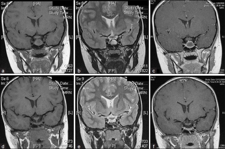
Figure 1.

(a–c) Coronal MR image showing a cystic lesion in the middle of the pituitary gland, measuring 17.1 × 13.9 × 11.4 mm3. (a) On T1-weighted imaging, the lesion is mostly isointense to brain, with a small hyperintense intracystic nodule. (b) On T2-weighted imaging, the lesion is hyperintense to brain, with the intracystic nodule being hypointense. There is a concentric hypointense region surrounding the lesion. (c) Contrast-enhanced T1-weighted imaging shows peripheral enhancement. (d–f) Approximately 1 year later, the size of the lesion is dramatically decreased. (d) This T1-weighted image shows a flattened gland that is approximately 3 mm in height. (e) T2-weighted image similarly demonstrates a flattened gland with no definite mass. A hypointense rim surrounds the gland. (f) T1-weighted image with contrast demonstrates no abnormal enhancement.
