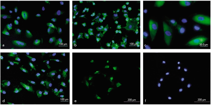
Fig. 3.
Immunohistochemical analyses of mesenchymal, neural and epithelial markers expression in cultured AF cells. Stained with antibodies against (a) CD105; (b) CD49d; (c) β3-tubulin; (d) keratin 19; (e) p63; (f) the same field, nuclei stained with DAPI. (a, b, c, d) Merge, nuclei stained with DAPI

