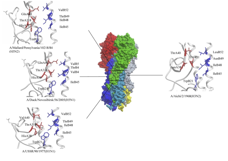
Fig. 7.
Conserved conformational epitopes of hemagglutinins recognized by neutralizing antibodies specific to the influenza strains used in this work and belonging to caldes Н1 and Н3. The surface of the hemagglutinin trimer is depicted in the middle; the HA1 chains are depicted in red, green and grey; and the HA2 chains are shown in dark blue, light blue and yellow. The region which contains the epitopes for the neutralizing antibodies is marked by a black box. The epitopes and the key amino acids that form them are depicted in HA1 (red) and HA2 (blue) hemagglutinin chains of various influenza viruses: A/Aichi/2/68(H3N2) (1EO8), A/USSR/90/77(H1N1) (1RVX), A/Mallard/Pensylvania/10218/84H5N2(H5N2) (1JSM) and A/Duck/Novosibirsk/56/2005(H5N1) (2IBX)

