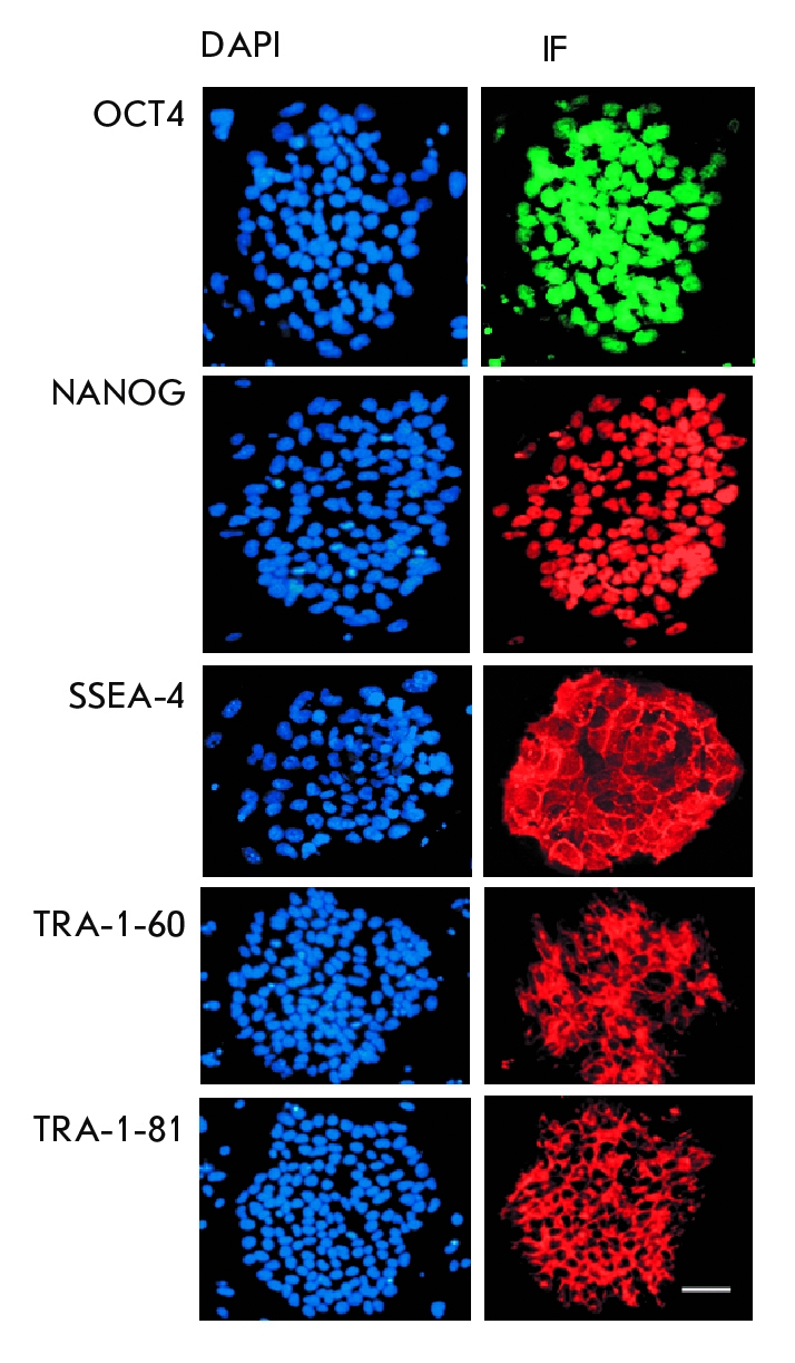
Fig. 3.

Immunocytochemical staining (IF) of iPSC colonies with antibodies against the transcription factors OCT4 (green) and NANOG (red) and against the surface antigens SSEA-4, TRA-1-60, and TRA-1-81 (red). Cell nuclei are counterstained with DAPI (blue). Scale 100 µm.
