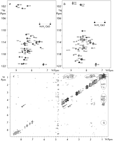
Fig. 1.
a and b – Heteronuclear NMR spectrum 1 H- 15 N HSQC of 15 N-labeled GpAtm in DPC micelles and DMPC/DHPC (1/4) bicelles, respectively, with a molar peptide/protein ratio of 1:35, at 40°C and pH 5.0. The resonance assignments are shown. c – The intermolecular proton-proton NOE contacts are presented on 2D project of the 3D 15 N, 13 C F1-filtered/F3-edited-NOESY spectrum acquired for the “isotopic-heterodimer” GpAtm sample embedded into the DPC micelles. The NOE cross peaks from the side chain hydroxyl ОγН group of T 87 are labeled by arrows, revealing the intramolecular hydrogen-bonding of the hydroxyl group with the carbonyl group of G 83 in major conformation of the GpAtm dimer.

