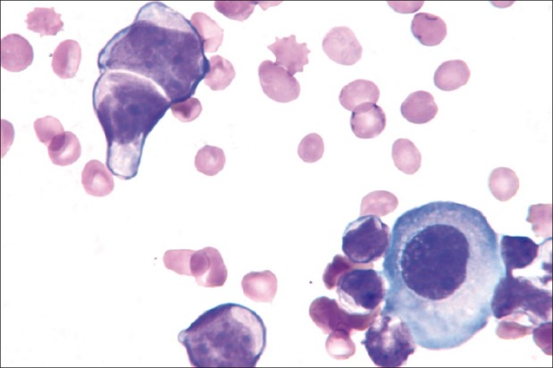
Figure 1.

A pair of neoplastic cells (upper left) shows scant cytoplasm, irregular nuclear contours, visible small nucleoli, and a possible inter-cellular ‘window’. The cytoplasmic eosinophilia of the neoplastic cells contrast with the cytoplasmic basophilia of the larger reactive mesothelial cell (lower right) (Wright stain, ×1000)
