Figure 2.

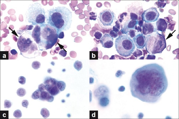
(a, b) Neoplastic cells (arrows) intermixed with reactive mesothelial cells. Note the eccentric location of the nuclei of neoplastic cells. (c) Tight 3D cluster of small undifferentiated tumor cells. (d) Multilobated / multinucleated, large, pleomorphic tumor cell with dense cytoplasm (a, b. Wright stain, c, d. Papanicolaou stain, all images original magnification, ×1000)
