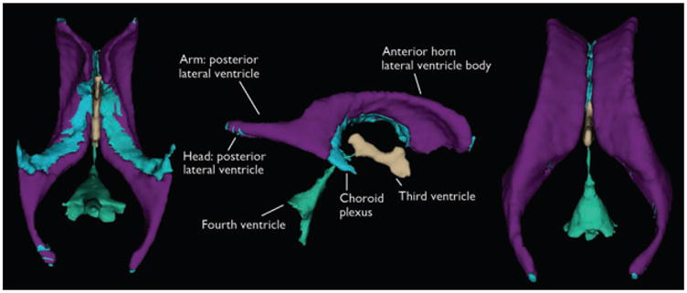
Figure 1.

Visualization of ventricular component segmentation produced from FreeSurfer for an individual participant. The views are from the ventral aspect with the anterior margin at the top (left view), lateral aspect with the anterior margin to the right (middle view), and dorsal aspect with the anterior margin at the top (right view).
Epilepsia © ILAE
