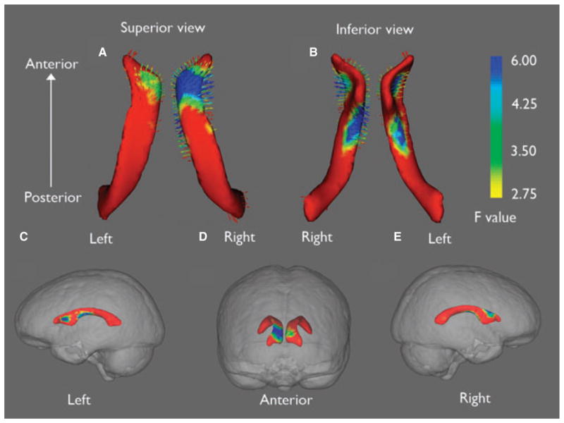
Figure 3.

Selective expansion of lateral ventricles in IGE. Shape analysis showed expansion (arrows pointing outward) of lateral ventricles is selective mainly for bilateral superior horns (A, B). Lateral ventricle shape differences are superimposed on glass brains to illustrate approximate brain regions surrounding areas of ventricular expansion (C, D, E). On the right side, ventricular enlargement is located in brain regions surrounding lateral and medial frontal lobe as well as basal ganglia (C, D). On the left side, the ventricular expansion is more localized to regions surrounding the medial frontal region and basal ganglia (D, E). Color bar indicates the statistic for the shape analysis, F (3,74) with associated probabilities of p = 0.12 (red), p = 0.02 (green), and p = 0.003 (blue).
Epilepsia © ILAE
