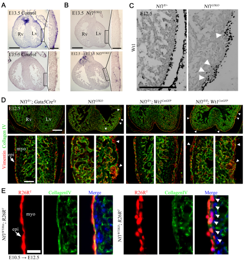
Fig. 1.
Disruption of epicardial development by loss of Nf1 in epicardium. (A,B) Nf1 mRNA expression was detected by in situ hybridization in heart sections of the indicated genotype. Nf1WTiKO mouse embryos were maternally induced with tamoxifen for Cre activity at E12.5 for 24 hours before processing (E12.5 → E13.5). The boxed regions are shown at higher magnification in the insets. (C) Immunohistochemistry (IHC) for the epicardial marker Wt1. Arrowheads indicate increased invasion of Wt1+ cells. (D) IHC for vimentin and collagen IV in embryonic hearts of the indicated genotype. Arrowheads indicate expansion of epicardial cells into the subepicardium. Bottom panels are higher magnifications of left and right ventricle. Arrow indicates epicardium. (E) R26RT fluorescence in heart sections of the indicated genotype. Oral tamoxifen administration is indicated by the stage of administration followed by the stage of isolation (E10.5 → E12.5). Arrows indicate epicardium. Arrowheads indicate migrated epicardial cells (below the basement membrane, collagen IV). Rv, right ventricle; Lv, left ventricle; epi, epicardium; myo, myocardium. Scale bars: 500 μm in A,B; 100 μm in A,B insets; 200 μm in C,D top; 50 μm in D bottom; 25 μm in E.

