
Figure 2.
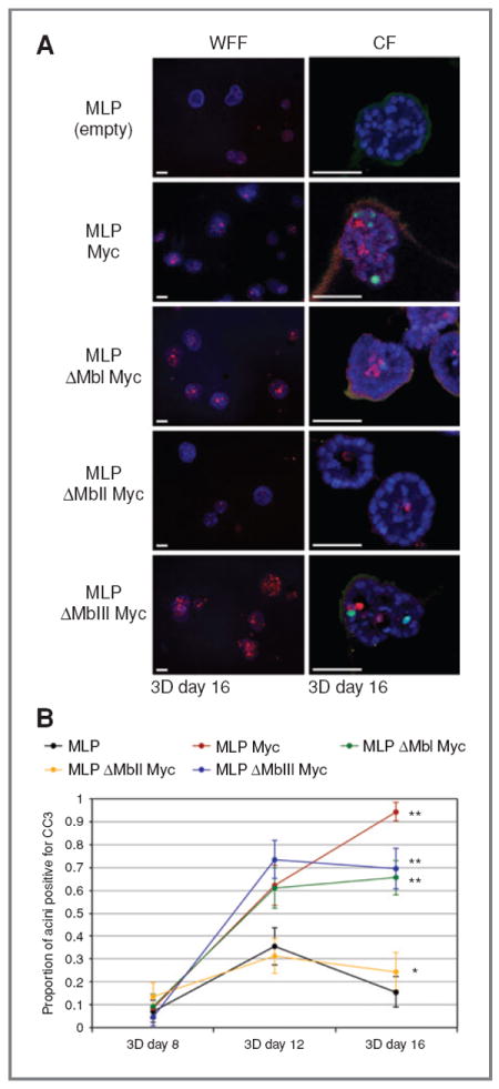
Myc-induced apoptosis in developing MCF10A acini requires MbII. A, wide-field fluorescence (WFF) and confocal (CF) images of CC3 (red, both WFF and CF) and Ki67 (green, CF only) immunofluorescence in day 16 acini expressing an empty vector (MLP), Myc (MLP Myc), a Myc box I deletion mutant (MLP ΔMbI Myc), a Myc box II deletion mutant (MLP ΔMbII Myc), or a Myc box III deletion mutant (MLP ΔMbIII Myc). Scale bars, 50 μm. B, quantification of the proportion of acini with 3 or more cells staining positive for CC3 at days 8, 12, and 16. Error bars, ±1 SEM for the population proportion (χ2 test compared with MLP: *, P = 0.10; **, P < 0.004).