Figure 5.

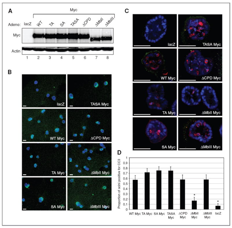
Myc induces apoptosis in mature 3D MCF10A acini. A, quantitative Western blot image showing adenoviral Myc expression relative to actin in mature 3D (day 20) acini (Supplementary Fig. S6A and B). Adenoviral vectors expressing lacZ, Myc (WT Myc), T58A Myc (TA Myc), S62A Myc (SA Myc), T58A S62A Myc (TASA Myc), a CPD-deletion mutant missing L56P57T58P59 (ΔCPD Myc), a Myc box II deletion mutant (ΔMbII Myc), or a Myc box III deletion mutant (ΔMbIII Myc) are shown. B, wide-field images showing adenoviral Myc immunofluorescence (green) in mature 3D (day 20) acini. Nuclei are counterstained with Hoechst 33342 (blue). C, confocal images of CC3 (red) and Ki67 immunofluorescence (green) in MCF10A acini in 3D culture at day 20 with adenoviral vectors from A. Scale bars (B and C), 50 μm. D, quantification of the proportion of acini with 3 or more cells staining positive for CC3. Error bars, ±1 SEM (*, P < 0.007, χ2 test compared with adenoviral WT Myc).
