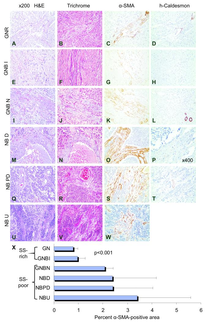
Fig. 1. Cancer Associated Fibroblasts in Schwannian Stroma-poor neuroblastoma tumors.
Representative sections of human ganglioneuroma[GNR] (A-D), ganglioneuroblastoma intermixed[GNBI] (E-H), ganglioneuroblastoma nodular[GNBN] (I-L), and neuroblastoma tumors that are differentiating[NBD] (M-P), poorly differentiated[NBPD] (Q-T), and undifferentiated[NBU] (U-W) tumors stained with H&E (A, E, I, M, Q, and U), Masson Trichrome special stain (B, F, J, N, R, and V), α-SMA which reveals cancer-associated fibroblasts and pericytes (C, G, K, O, S, and W), and high molecular weight caldesmon, which stains pericytes, but not cancer-associated fibroblasts (D, H, L, P, and T). The mean (±standard deviation) percent of α-SMA positive areas per total tumor area analyzed for each tumor type are shown in the bar graph (X). α-SMA positive, caldesmon-negative cancer-associated fibroblasts are rare in the Schwannian Stroma[SS]-dominant ganglioneuroma (C, D and X) and Schwannian Stroma -rich ganglioneuroblastoma intermixed (G/H and X) tumors. Significantly more cancer-associated fibroblasts are present in Schwannian Stroma -poor tumors (K, L, S, T, W). Bands of fibrocollagenous stroma are thin and delicate in ganglioneuroma and ganglioneuroblastoma intermixed tumors, but are thick and support microvascular proliferation in ganglioneuroblastoma nodular and neuroblastoma tumors (pale blue B and F vs. J, N, R and V). Cancer-associated fibroblasts constitute the majority of stromal cells within fibrovascular stroma (O, S and W). Original magnification x200 (A-L, T) and x400 (P).

