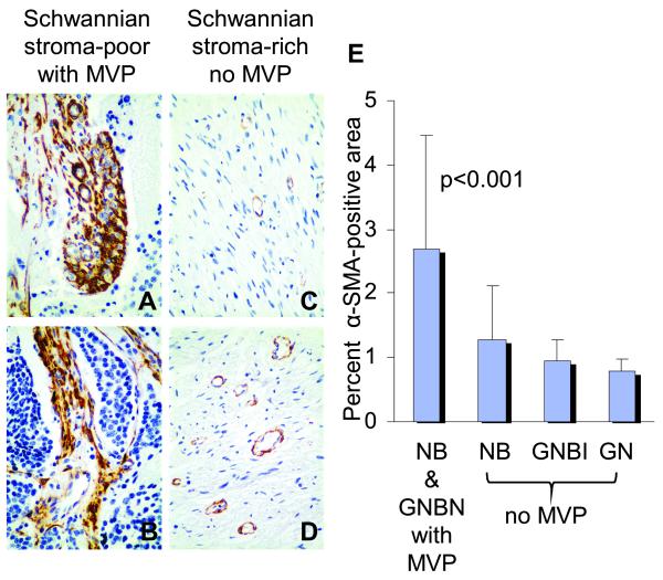
Fig. 2. Cancer-associated fibroblasts in human neuroblastomas with microvascular proliferation.
Representative sections of differentiating neuroblastoma[NBD] (A), poorly differentiated neuroblastoma[NBPD] (B), ganglioneuroma[GNR] (C) and ganglioneuroblastoma intermixed[GNBI] (D) tumors immunostained for α-SMA. The mean (±standard deviation) percent of α-SMA -positive areas per total tumor areas analyzed for tumors with microvascular proliferation[MVP] are shown in the bar graph (E). Significantly more α-SMA -positive cancer-associated fibroblasts are present in the tumors with microvascular proliferation (A, B, and E) as compared to tumors without microvascular proliferation (C, D, and E) (p<0.001). The cancer-associated fibroblasts are present within bands of fibrovascular stroma supporting microvascular proliferation (A and B). Original magnification x400.

