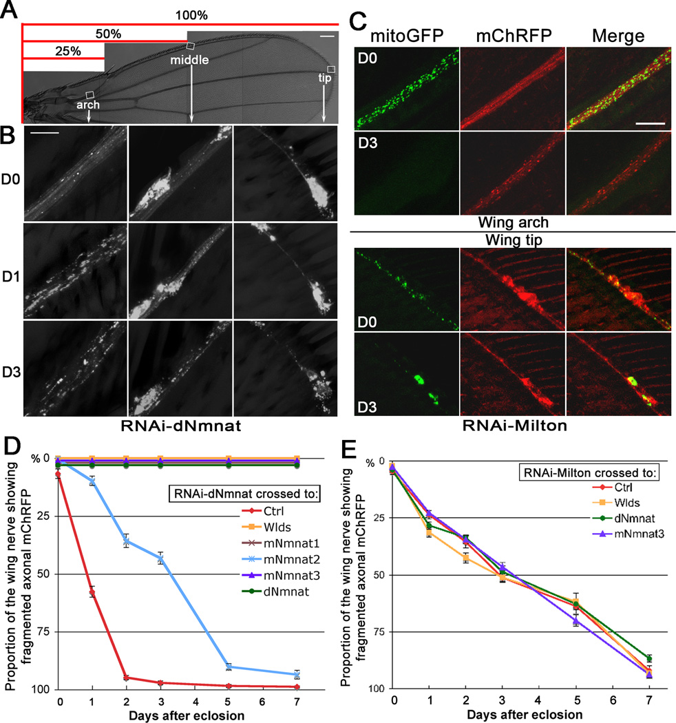
Figure 3. Knockdown of Nmnat or Milton in the wing nerve induces spontaneous axon degeneration.
(A–B) dpr>RNAi-dNmnat flies exhibited retrograde, spontaneous axon degeneration. Fragmentation of axonal mChRFP was seen earlier and more prominent in the wing arch (distal axons) than in the wing tip (proximal axons). (C) dpr>RNAi-Milton flies had normally distributed mitoGFP and smooth axonal mChRFP on D0. On D3, mitoGFP was depleted from the axons (wing arch) and retained in the cell bodies (wing tip); axonal mChRFP was massively fragmented in the wing arch but remained continuous in the wing tip. (D–E) Degeneration curves of dpr>RNAi-dNmnat (D) and dpr>RNAi-Milton (E) flies, plotted as proportion of fragmented wing nerve (see the scale in (A)). Ctrl, UAS-Luciferase. Mean ± SEM is shown, n = 14~42. Scale bar: 100 µm in (A), 10 µm in (B), and 20 µm in (C).

