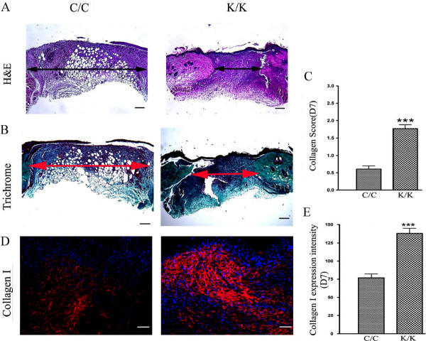
Figure 3.
Histological analysis of PPARγ KO (K/K) mice and WT (C/C) wound tissue. (A) H&E and (B) trichrome staining of day 7 wounds (C/C: N = 10; K/K: N = 12, original magnification × 5, bar = 200 μm). The arrow indicates wound width. (C) The collagen content in each section was assessed by three blinded observers using the following assessment criteria: 0 signifies no collagen fibers, 1 signifies a few collagen fibers, 2 signifies a moderate amount of collagen fibers, and 3 signifies an excessive amount of collagen fibers. (D) Immunofluorescence analysis using an anti-type I collagen antibody. (WT: N = 10; KO: N = 12, original magnification × 20, bar = 50 μm). Red-PPARγ; Blue-DAPI. (E) Quantification of type collagen I protein expression in wound tissues at Day 7 after wounding. *** = significant difference between C/C-WT and K/K-KO groups (P < 0.001). DAPI, 4',6-diamidino-2-phenylindole; PPARγ, peroxisome proliferator-activated receptor-γ Loss of PPARγ results in enhanced collagen, α-SMA, PCNA and CCN2 expression.

