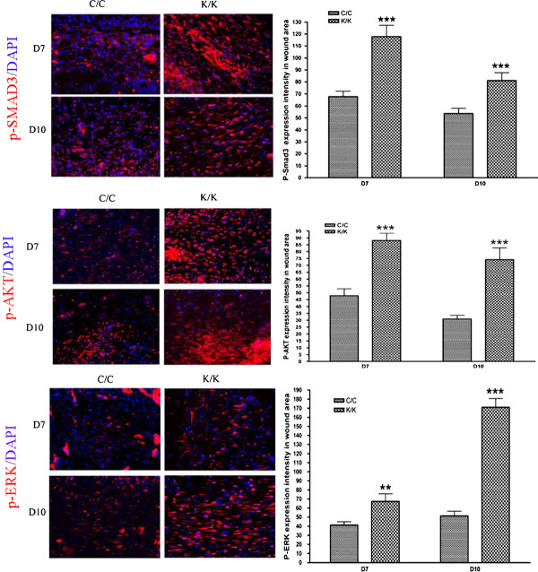
Figure 6.
Elevated p-Akt, p-Smad3 and p-Erk staining in PPARγ KO (K/K) wound tissue (day 7 and day 10 post-wounding). Indirect immunofluorescence analysis with anti-, anti-p-Smad3 and anti-p-Erk antibodies, as indicated, in wound tissues day7 and day 10 post-wounding (C/C: N = 10; K/K: N = 12, original magnification × 20, bar = 50 μm). p-Akt, p-Smad3 and p-Erk expression intensity, respectively, are shown. Asterisks indicate a significant difference between C/C and K/K groups (** = P < 0.01; *** = P < 0.001). PPAR, peroxisome proliferator-activated receptor-γ.

