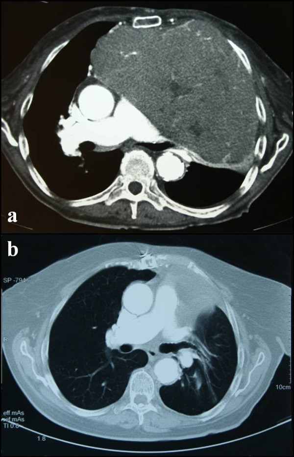
Figure 2.

Pre-operative and post-operative chest computed tomography scans. (a) Pre-operative chest computed tomography scan reveals a soft-tissue-density lesion of the anterior mediastinum. (b) Post-operative chest computed tomography scan (same level imaging sections), performed two years after resection of the tumor, shows: no evidence of tumor, the esophagus returned to its anatomical position, decompression of the left main bronchus, expansion of the left lung, correction of the mediastinal width and regain of chest wall subcutaneous fat tissue.
