Figure 2.
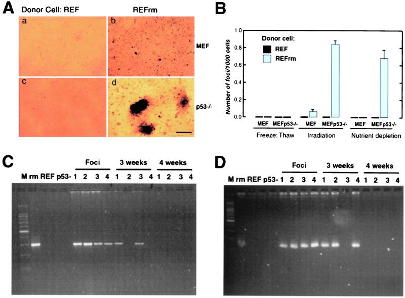
Apoptotic bodies derived from REFrm cells induce focus formation in MEF p53−/− cells. (A) MEF (a and b) or MEF p53−/− cells (c and d) were cocultured with either apoptotic REF (a and c) or REFrm (b and d) and focus formation was analyzed after 8 days in culture. (Bar = 220 μm.) (B) Frequency of focus formation in MEF and MEFp53−/− cells after coculture with necrotic cells or apoptotic REF or REFrm cells. Necrosis was induced by freeze thawing, and apoptosis was induced either by irradiation or by nutrient depletion, as indicated. Results are shown as mean ± SD of three independent experiments (C) and (D). PCR analysis for the presence of H-rasV12 (C) and human c-myc (D) in donor REFrm (rm), REF, recipient MEF p53−/− (p53-), foci (1–4), and the resulting cell lines after 3 and 4 weeks of propagation. M = 100-bp DNA ladder.

