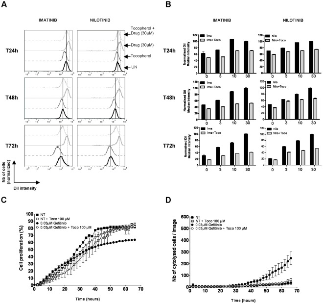
Figure 5. α-tocopherol prevents the action of kinase inhibitors used in clinical practice. A, B.
DiI-stained K562 cells were treated with increasing amounts of imatinib or nilotinib (0, 3, 10 or 30 µM) supplemented with vehicle or 1 µM of α-tocopherol. In each condition, cell proliferation was followed according to the intensity of DiI staining after 24, 48 and 72 hours. Cells exhibiting a brighter DiI fluorescence than control cells divide more slowly and are delayed in their cycle cell progression, while the ones having an equal or a weaker DiI intensity proliferate at the same rate or faster than control cells. An example of DiI fluorescence intensity post-imatinib or nilotinib treatment (30 µM) in presence or absence of 1 mM α-tocopherol is shown at each time point in A. The average DiI fluorescence intensity after all treatment is reported in panel B after normalization (100% correspond to the DiI most intense cells). C PC-9 cells were treated with DMSO or 0.03 µM of Gefitinib in presence or absence of 100 µM of α-tocopherol and immediately transferred to the IncuCyte system for kinetic cell growth measurement. The percentage of cell growth corresponds to the surface occupied by the cells in each well at each time point. D PC-9 cells were treated with DMSO or 0.03 µM of Gefitinib in presence or not of 100 µM of α-tocopherol, and incubated in presence of SYTOX Green in the IncuCyte system for cell death measurement.

