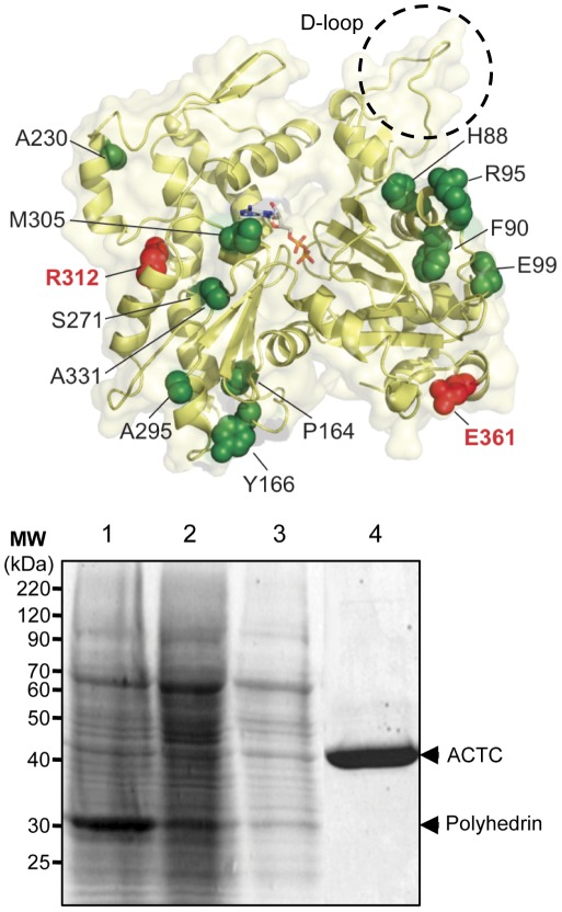
Figure 1. Cardiac actin mutations related to cardiomyopathies.
A . The protein structure of actin (PDB 1J6Z) [39] showing the locations of mutations related to HCM (in green spacefilling) or DCM (in red spacefilling) and bound ATP (sticks) visualized using PyMol [40]. The D-loop of actin is highlighted with a dashed circle. ACTC substitution mutations related to HCM shown are: H88Y, F90del, R95C, E99K, P164A, Y166C, A230V, S271F, A295S, R312C, A331P, and M305L. The two ACTC mutants associated with DCM are R312H and E361G. The amino acid numbers listed are based on the sequence of the protein after posttranslational processing of the N-terminus, which removes the first two amino acids of the nascent polypeptide. Note that mutations at Arg-312 are associated with both HCM and DCM. B . Purification of baculovirus expressed ACTC mutant protein. Sf9 cells were infected with recombinant baculovirus expressing the A230V ACTC mutant protein at an MOI of 1 for 72 hours. Shown is a 10% SDS-PAGE of samples taken throughout the purification: lane 1, crude lysate; lane 2, supernatant fraction of lysates; lane 3, lysate following filtration and applied to a DNase-I affinity column; lane 4, purified A230V ACTC protein eluted from the DNase-I affinity column. Arrows indicate the position of actin and the polyhedrin protein expressed by the baculovirus in Sf9 cells.

