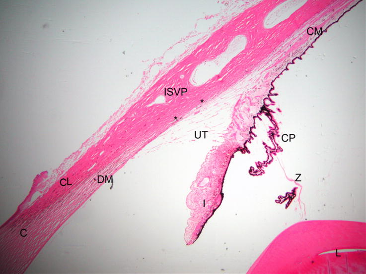
Figure 1.

Photomicrograph illustrating the morphology of the normal feline outflow pathway. (C = cornea, DM = Descemet’s membrane, CL= corneoscleral limbus, I = iris, L= lens, CP = ciliary processes, ISVP = intrascleral venous plexus, asterisks mark angular aqueous plexus, UT = uveal trabecular meshwork)
