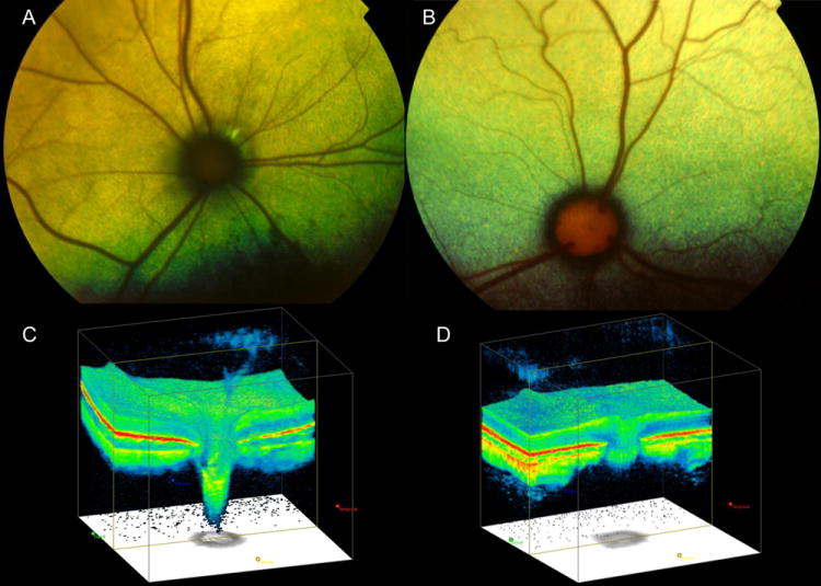
Figure 4.

Fundus photographs illustrating (A) the optic nerve head (ONH) appearance of a cat with advanced primary congenital glaucoma, with ONH cupping and optic nerve degenerationand (B) a normal cat, which demonstrates normal prominence of the laminar pores. Note that in the glaucomatous cat (A) the ONH is small and dark and surrounded by a dark ring and by focal peri-papillary hyper-reflectivity. Optic nerve cube scans acquired by spectral-domain Optical Coherence Tomography (OCT; Cirrus, Carl Zeiss Meditec Inc., Dublin, CA) in a cat with glaucoma that demonstrates dramatic posterior displacement of the lamina cribrosa (C) compared to a normal cat (D).
