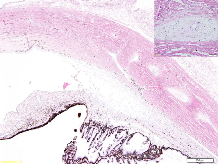
Figure 6.

Photomicrograph illustrating myxomatous changes surrounding the intrascleral veins (inset) in a cat with primary open angle glaucoma. The ciliary cleft is open and the trabecular meshwork appears normal. (Image courtesy of Dr. R.R. Dubielzig, Comparative Ophthalmic Pathology Laboratory of Wisconsin)
