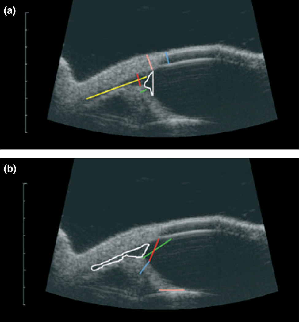
Figure 1. High-resolution ultrasound images of a normal feline eye with measurement parameters indicated.
Scale bar to left of each image indicates millimeters. (a) ARA = angle recess area, white; ST = scleral thickness, pink; CT = corneal thickness, blue; IT = iris thickness, green; MLCC = maximum length of ciliary cleft, yellow; MWCC = maximum width of ciliary cleft, red; and (b) ACC = area of ciliary cleft, white; TMID = trabecular meshwork-iris distance, red; ICPD = iridociliary process distance, red/blue; AOD500 µm = angle opening distance at 500 µm, green; ILC = iris lens contact, pink.

