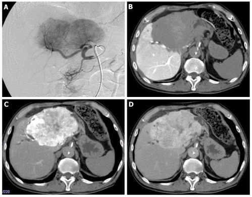
Figure 2.
Follow-up hepatic arterial angiography and computed tomography images after 7 years. A: Hepatic arterial angiography showed hypervascular tumor of left liver lobe; B: Computed tomography (CT) angiography (portal phase) showed portal vascularity defect in the tumor; C-D: CT angiography (C: Arterial early phase; D: Arterial late phase) showed hypervascularity and a persistent enhancement effect.

