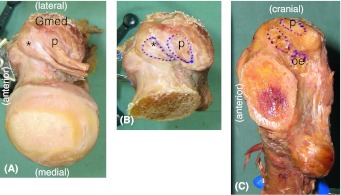
Fig. 2A–C.
The attachment site of the short external rotator muscles indicates their relative localization. (A) A superior view, (B) superior view and footprint of the tendon insertion, and (C) mediolateral view and footprint of the tendon insertion are shown. *Conjoined tendon of the gemellus superior, obturator internus, and gemellus inferior; p = piriformis; oe = obturator externus; Gmed = gluteus medius.

