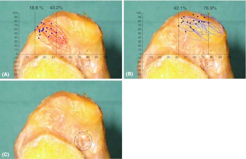
Fig. 4A–C.
Superimposed footprints of the attachment of the short external rotator muscles on the inner aspect of the greater trochanter are shown. (A) The attachment of the conjoined tendon (obturator internus, gemellus superior, and gemellus inferior) for 20 hips is shown. The plot highlights the most anterior part of the attachment in each specimen to show its variation of 18.8% to 43.2% from the anterior border of the greater trochanter. (B) The attachment of the piriformis for 20 hips is shown. The plot highlights the most anterior part of the attachment in each specimen. (C) The dotted line indicates the attachment site of the obturator externus to a fossa.

