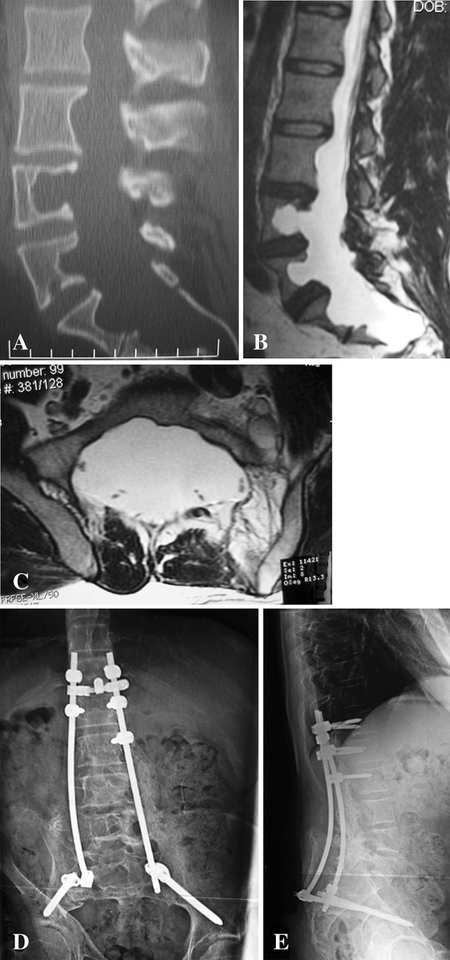
Fig. 2A–E.

(A) A sagittal CT scan shows the spine of a 29-year-old man with neurofibromatosis and multilevel duralectasia who had presented with severe disabling pain with ambulation. (B) A sagittal T2-weighted MR image demonstrates duralectasia with erosion of the middle column of the L3, L4, L5, S1, and S2 vertebral bodies. (C) An axial T2-weighted MR image at S1 demonstrates severe duralectasia. (D) An AP plain radiograph shows the spine after posterior spinal fusion with instrumentation from T12 to the pelvis. Postoperative prophylactic bed rest was prescribed for 3 months due to the limited fixation, high risk of pseudarthrosis, and limited treatment options in the event of failure of this procedure. (E) A lateral plain radiograph at last followup when he was ambulating without assistive devices and plain radiographs showed maintenance of alignment without fractures of the bone, failure of instrumentation, or failure of fixation of the instrumentation.
