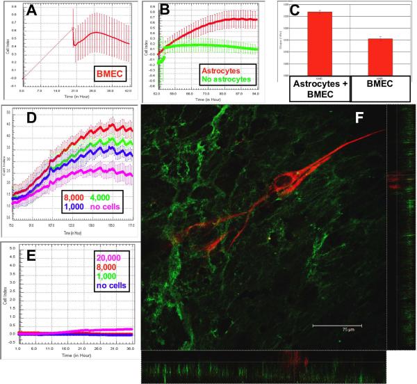
Figure 2. BMEC adhesion is increased following astrocyte coincubation.
The Cell Index (CI) increases during the adhesion of BMEC to the inverted filter assembly. When the plate is reverted and connected to the xCELLigence system, it is apparent that BMEC have adhered and begun to spread (A). When astrocytes are added to the upper well, the CI is increased further compared to BMEC monocultures (B, p<0.0001). The change in CI is calculated using the installed software when astrocytes (C, left trace) are added to the wells. Astrocytes increased the CI in a dose-dependent manner (D). Astrocyte foot processes did not cross the filter and increase the CI in the absence of BMEC (E). Endothelial cells were immuno-labeled with antibodies to Von Willebrand Factor (F, green). Astrocytes were labeled with antibodies to GFAP-CY3 (red). Monolayers of endothelial cells were apparent below the GFAP-positive astrocyte processes. Confocal stacks were then examined in x–z and y–z format to definitively show that the astrocytes were physically separated from the endothelial cells (see also the supplementary video).

