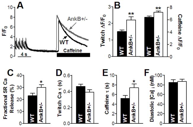
Figure 1. Larger Ca transients and SR Ca content and reduced NCX function in myocytes from AnkB+/− mice.
(A) Ca transient and SR Ca content measurements - representative traces in a WT and an AnkB+/− myocyte. Myocytes were paced at 1 Hz until Ca transients reached steady-state, then pacing was stopped for 10 sec, followed by application of 10 mM caffeine. Mean values for Ca transient amplitude and SR Ca content (18 cells, 5 animals for both WT and AnkB+/− mice) (B), SR fractional release (18 cells, 5 mice for both WT and AnkB+/− mice) (C), decay time of the twitch Ca transient (18 cells, 5 mice for both WT and AnkB+/− mice) (D) and decay time of the caffeine-induced Ca transient (9 cells, 5 mice for both WT and AnkB+/− mice) (E). (F) Mean diastolic [Ca]i in myocytes from WT and AnkB+/− mice (n=10, 4 mice for both WT and AnkB+/− mice).

