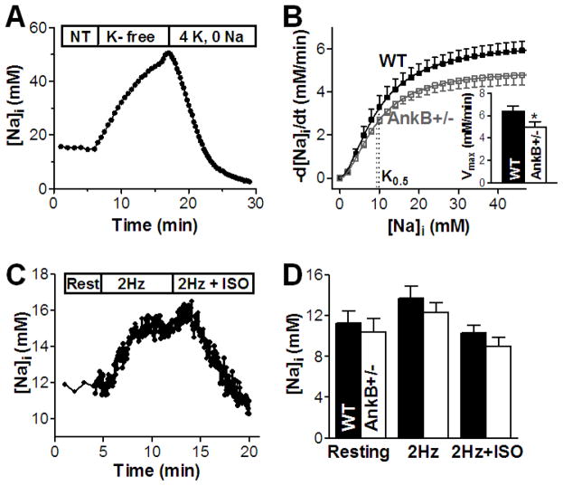
Figure 2. NKA function is reduced but [Na]i is unchanged in myocytes from AnkB+/− mice.
(A) Measurement of NKA-mediated Na efflux in intact myocytes from WT and AnkB+/− mice - representative trace in an AnkB+/− myocyte. NT – normal Tyrode’s solution; (B) The rate of [Na]i decline (−d[Na]i/dt) was calculated by numerical differentiation, plotted as a function of [Na]i and fitted with Hill equation to determine Vmax, K0.5 and the Hill coefficient. Mean data in myocytes from WT (8 cells, 6 mice) and AnkB+/− mice (10 cells, 5 mice) indicate that K0.5 is not changed whereas Vmax is significantly lower in AnkB+/− myocytes (Inset). (C) [Na]i measurements at rest and during pacing (2Hz) in the absence and in the presence (2Hz+ISO) of 1 μM isoproterenol in a WT myocyte. (D) Mean [Na]i in myocytes from WT (12 cells, 7 mice) and AnkB+/− mice (14 cells, 6 mice) at rest and during pacing with and without isoproterenol.

