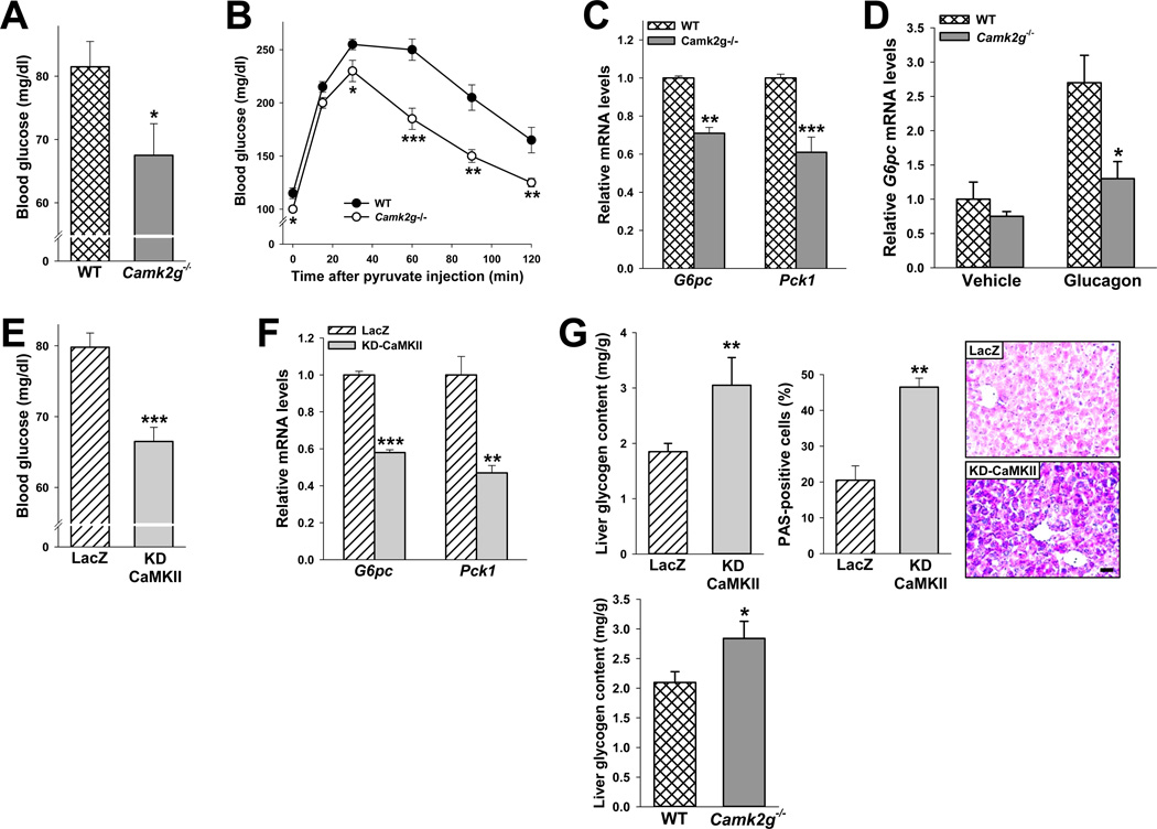
Figure 3. CaMKIIγ deficiency or acute inhibition in vivo decreases blood glucose and hepatic G6pc and Pck1.
(A) Blood glucose of 12-h-fasted 8-wk/o WT and Camk2g−/− mice (*P < 0.05). (B) As in (A), but the mice were fasted for 18 h and then challenged with 2 mg kg−1 pyruvate (B) (*P < 0.05; **P < 0.01; ***P < 0.005; mean ± S.E.M.). (C) Liver G6pc and Pck1 mRNA in 12-h-fasted WT and Camk2g−/− mice (**P < 0.01; ***P < 0.001; mean ± S.E.M.). (D) WT and Camk2g−/− mice were injected i.p. with glucagon (200 µg kg−1) and sacrificed 30 min later. Liver G6pc mRNA was assayed (*P < 0.05; mean ± S.E.M.). (E–G) 9-wk/o WT mice were administered 1.5 × 10⁹ pfu of adeno-LacZ or KD-CaMKII, and 5 days later the following parameters were assayed in 12-h-fasted mice: E, blood glucose (***P < 0.001; mean ± S.E.M.); F, liver G6pc and Pck1 mRNA (**P < 0.01; ***P < 0.001; mean ± S.E.M.); and G, liver glycogen content and PAS-positive cells (**P < 0.01; mean ± S.E.M.). Panel G also shows liver glycogen content in fasted WT and Camk2g−/− mice (*P < 0.05; mean ± S.E.M.). For all panels, n = 5/group except panel D, where n = 4/group.

