Figure 3.
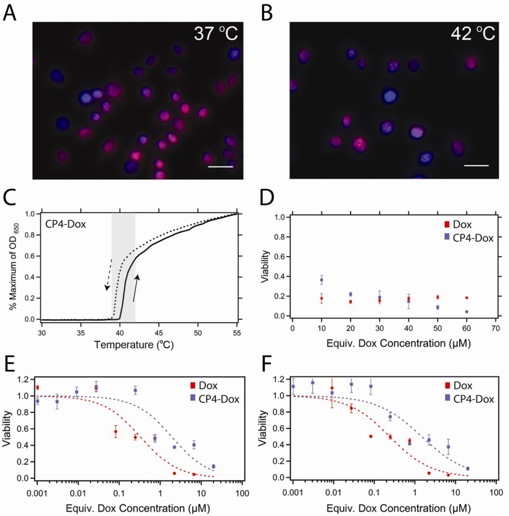
In vitro cytotoxicity of CP-Dox nanoparticles. (A) Fluorescence microscopy of C26 cells following 1 h incubation at 37 °C or (B) 42 °C with CP-Dox nanoparticles at 5.6 µM (30 µM Dox equivalents). Colocalization of Dox (red) with Hoechst stain (blue) suggests that the Dox localizes to the nucleus with and without the application of heat. Scale bars indicate 25 µm. (C) The phase transition of CP4-Dox is fully reversible in complete cell media. CP4-Dox transitions within the hyperthermia window (shaded) at 40.2 °C at 25 µM CP. (D) Viability of C26 cells after mild hyperthermia (1 h at 42 °C) and 24 h incubation with Dox at 37 °C (red) or CP-Dox nanoparticles (blue). (E) Viability of C26 cells following a 1 h exposure at 37 °C or (F) 42 °C and a 72 h incubation in Dox (red) or CP-Dox nanoparticles (blue). Concentrations below 0.1 µM likely result in the disassembly of the nanoparticle system.

