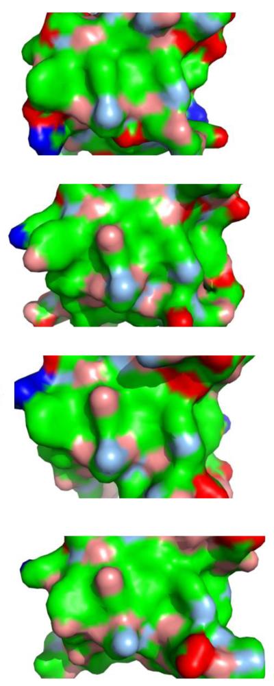
Figure 2.
Ligand binding surfaces of SH3 domains. Despite belonging to different proteins, and despite being implicated in different signaling pathways[sta12], the molecular characteristics of SH3 ligand binding surfaces are highly similar. From top to bottom:[sta13] CIN85 (PDB id 2BZ8), Fyn (1SHF), Grb2 C-terminal (2W0Z), Abl (1BBZ). Surfaces are colored according to blue, positively charged atoms; red, negatively charged atoms; green, hydrophobic atoms; salmon, polar oxygens; marine, polar nitrogens; yellow, sulfur.

