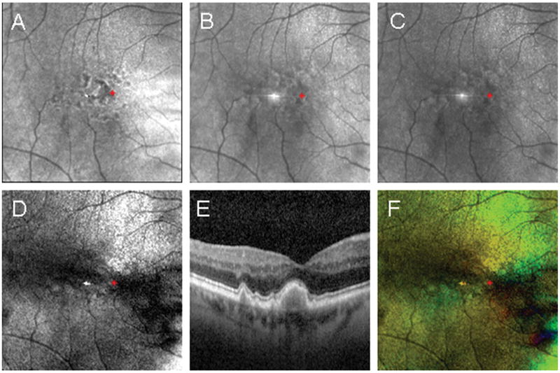
Figure 4.
Example of foveal localization in a subject with large coalesced drusen in the central macula confirmed using spectral domain optical coherence tomography. The red asterisk indicates the foveal location determined using the Maximum Phase of the Crossed Detector Image. Foveal coordinates were then plotted on the remaining images derived from the same raw data set. (A) The Depolarized Light Image, with the least polarization content, emphasizes the pathology in the outer retina. (B) The Confocal Image and (C) The Parallel Detector Image, do not have features that appreciably aid in the localization of the fovea. (D) The Birefringence Image contains only amplitude information represented in grayscale, but the macular cross pattern is disrupted and does not provide optimal information for foveal localization. (E) The SDOCT image shows the location of pathological features emphasized in the Depolarized Light Image. (F) The Maximum Phase of the Crossed Detector Image, color code as above, again clearly demarcates the fovea center despite central macular pathology.

