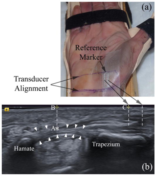
Figure 1.
A cadaveric hand is secured in a custom-made splint (a) for ultrasound imaging (b). White arrowheads indicate the TCL. Points A (‘*’), B (‘+’ on the left), and C (‘+’ on the right) indicate the thenar muscle’s ulnar point (TUP), the surface projection of the TUP, and the surface intersection of the left interference line generated by the reference marker, respectively.

