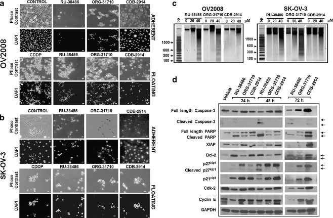
Fig. 5.
Lethality of antiprogestins towards ovarian cancer cells. OV2008 or SK-OV-3 cells were cultured in the presence of 40 μM RU-38486, ORG-31710 or CDB-2914. After 60 h (OV2008) or 120 h (SK-OV-3), floating cells were collected, adhered to a microscope slide, stained with DAPI, and microscopic phase contrast and fluorescence images were obtained from the same fields. As positive control of apoptotic cell death, floating cells from cisplatin (CDDP) treated cells were included in the morphological studies a and b. Adherent untreated cells are also shown (CONTROL). Scale bar, 20 μm. c A similar experiment was done in which all floating and attached cells were pelleted, total DNA was isolated, subjected to agarose electrophoresis, stained with SYBR Gold nucleic acid stain, and photographed with the Amersham Typhoon fluorescence imaging system. A 100 base pair (bp) marker was run in parallel. (d) OV2008 cells were exposed to DMSO (Vehicle) or 40 μM RU-38486, ORG-31710 or CDB-2914 for 72 h. Whole protein extracts were obtained and separated by electrophoresis, and immunoblots were probed with the indicated antibodies to recognize cell cycle and cell death related proteins. The housekeeping gene GAPDH was used as protein loading control

