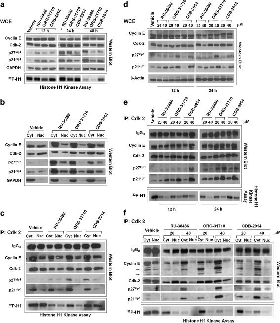
Fig. 6.
Effect of antiprogestins on cell cycle regulatory proteins in ovarian cancer cells. OV2008 cells were exposed to DMSO (Vehicle), 20 a-e or 40 μM d and e RU-38486, ORG-31710, or CDB-2914 for the indicated times a, d, and e or 48 h b and c. a Whole protein extracts (WCE) were obtained and separated by electrophoresis, and immunoblots were probed with the indicated cell cycle related antibodies. Whole protein extracts were also immunoprecipitated with anti-Cdk-2 antibody and assayed for their capacity to phosphorylate histone H1 in vitro in the presence of 32P ATP. b Isolation of nuclear and cytosolic fractions was achieved, proteins from each fraction were obtained and separated by electrophoresis, and immunoblots were probed with the indicated antibodies. c Nuclear and cytosolic extracts were imunoprecipitated with anti-Cdk-2 antibody, electrophoresed, and probed with the indicated antibodies. The immunoprecipitates were also assayed for their capacity to phosphorylate histone H1 in vitro in the presence of 32P ATP. d Time-course experiment on the effect of 20 or 40 μM antiprogestins on the expression of cell cycle- related proteins. e Whole cell extracts from previous experiment were imunoprecipitated with anti-Cdk-2 antibody, electrophoresed, probed with the indicated antibodies, and also assayed for their capacity to phosphorylate histone H1 in vitro. f A similar experiment was performed as in d but instead of WCE, nuclear and cytoplasmic protein extracts were isolated and immunoprecipitated with Cdk-2 antibody upon treatment of the cells with 20 or 40 μM antiprogestins for 24 h

