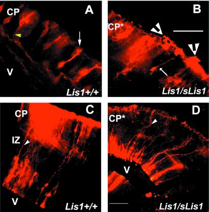
Figure 4.
DiI labeling of the cortex. (A and B) E14.5 DiI labeling of cortical cells. (Bar = 1 mm.) The white arrow labels the apical dendrite, and the yellow arrowhead marks the projecting axon. Lis1+/+ (A); Lis1/sLis1, white arrow marks a neuron that has “normal” morphology, and white arrowheads mark clusters of neurons that look abnormal (B). (C and D) E15.5 DiI labeling of cortical cells. (Bar = 25 μm.) Radial glia fibers are marked with arrowheads. Lis1+/+ (E), Lis1/sLis1 (F). V, ventricle; IZ, intermediate zone; CP*, abnormal CP.

