
Figure 1.
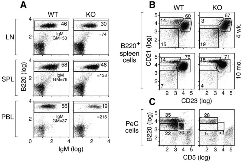
Altered B cell profiles in IκBNS KO mice. A) IκBNS KO mice exhibit a reduction in the percentage of B220+ B cells in the periphery. Cells prepared from the lymph nodes, spleens or blood of 9 month old C57BL/6 or IκBNS KO mice were examined for B220 and IgM surface expression by FACS. Numbers in boxes indicate the percentage of B220-positive cells, and the mean fluorescent intensity of IgM-FITC on B220+ cells is given for each sample. B) Abnormal generation of marginal zone B cells in IκBNS KO mice. B220+ splenic B cells from 4-week or 10-month old WT and IκBNS KO mice were examined for CD21 and CD23 by flow cytometry. Young IκBNS KO mice show a reduced percentage of CD21hiCD23lo marginal zone B cells. C) IκBNS KO mice lack B1 B cells in the peritoneal cavity. Peritoneal cavity cells isolated from WT or IκBNS KO mice were analyzed for B220+ CD5+ B cells. Gates show B2 (CD5-B220hi), B1a (CD5+B220int/lo) and B1b (CD5-B220lo) cells. B1a and B1b cell populations are absent from the IκBNS KO mouse peritoneal cavity. Three age-matched animals were examined in each set of experiments.